Left lateral decubitus position x ray – Left lateral decubitus position x-ray, a cornerstone of diagnostic imaging, offers unparalleled insights into the intricacies of the human body. This technique, meticulously executed, empowers clinicians with a wealth of information to unravel complex medical conditions.
Delving into the depths of this specialized imaging modality, we embark on a journey to explore its technical intricacies, clinical applications, and the spectrum of pathological findings it unveils.
Patient Positioning

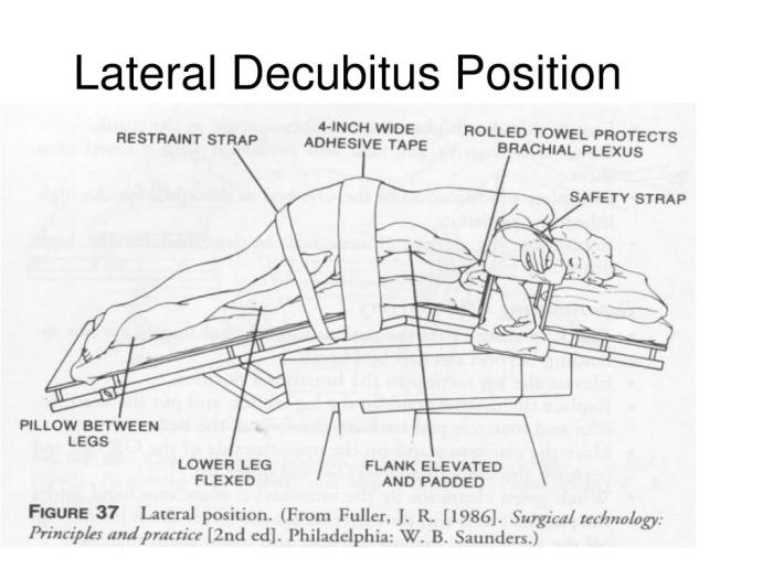
The left lateral decubitus position is a patient positioning technique used in medical imaging, particularly in X-rays, to visualize the thoracic and abdominal structures from the left side.
To position the patient correctly, follow these steps:
Patient Positioning
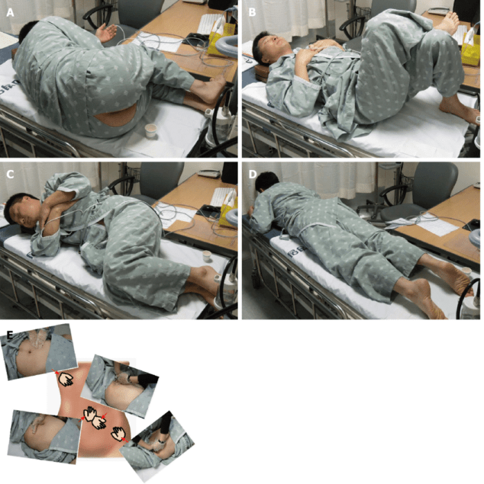
- Instruct the patient to lie on their left side with their right arm extended forward and their left arm behind their head.
- Ensure the patient’s head is supported by a pillow or foam pad to maintain a neutral position.
- Position the patient’s legs slightly flexed at the knees and hips to promote comfort.
- Use pillows or foam pads to support the patient’s body and prevent pressure points.
Radiographic Technique

To obtain optimal radiographic images in the left lateral decubitus position, it is crucial to employ appropriate radiographic techniques. These techniques involve selecting the optimal kVp, mAs, and beam angulation to ensure adequate penetration and image quality.
kVp and mAs, Left lateral decubitus position x ray
The selection of kVp and mAs depends on the patient’s size and the specific anatomical region being examined. Generally, higher kVp values are used for larger patients to achieve adequate penetration, while lower kVp values are preferred for smaller patients to minimize radiation exposure.
The mAs should be adjusted accordingly to ensure optimal image density and contrast.
Beam Angulation
The beam angulation in the left lateral decubitus position is typically adjusted to align with the anatomical structure of interest. This involves angling the beam perpendicular to the plane of the structure to minimize distortion and optimize image quality.
Anatomic Landmarks

The left lateral decubitus position provides a unique perspective of the thoracic and abdominal anatomy, allowing visualization of specific anatomic landmarks. Understanding these landmarks is crucial for accurate interpretation of radiographs obtained in this position.
Key Anatomic Landmarks
The following table summarizes the key anatomic landmarks visible in the left lateral decubitus position, along with their corresponding annotations:
| Landmark | Annotation |
|---|---|
| Heart | Opacifies the left hemithorax and is shifted posteriorly. |
| Aorta | Visible as a soft tissue density along the left lateral border of the heart. |
| Pulmonary artery | Projects anteriorly to the aorta and is seen as a curvilinear opacity. |
| Left main bronchus | Arises from the trachea and descends inferiorly and posteriorly. |
| Stomach | Contains gas and fluid levels and is located in the left upper quadrant. |
| Liver | Extends from the right hypochondrium to the left upper quadrant, below the diaphragm. |
| Spleen | Located in the left upper quadrant, posterior to the stomach. |
| Left kidney | Visualized as a bean-shaped opacity in the left upper quadrant. |
| Left adrenal gland | Lies superior to the left kidney. |
Pathological Findings: Left Lateral Decubitus Position X Ray
The left lateral decubitus position allows for the detection of various pathological findings within the chest and abdomen. These findings can provide valuable information for diagnosing and managing a range of conditions.
Common pathological findings that can be identified in the left lateral decubitus position include:
- Pleural effusions:Fluid accumulation in the pleural space, often associated with conditions such as heart failure, pneumonia, and pleural effusion.
- Pneumonia:Consolidation of lung tissue due to infection or inflammation, often characterized by increased density and air bronchograms.
- Lung abscesses:Collections of pus within the lung parenchyma, typically appearing as round or oval opacities with an air-fluid level.
- Interstitial lung disease:A group of conditions characterized by inflammation and fibrosis of the lung tissue, often resulting in reticular or ground-glass opacities.
- Mediastinal masses:Enlargement of mediastinal structures, such as lymph nodes, thymus, or vascular abnormalities, can be detected as soft tissue densities within the mediastinum.
- Diaphragmatic hernia:Protrusion of abdominal contents through an opening in the diaphragm, visible as gas-filled loops within the thoracic cavity.
- Ascites:Accumulation of fluid in the peritoneal cavity, often associated with liver disease, heart failure, or peritoneal inflammation.
- Hepatomegaly:Enlargement of the liver, which may be due to conditions such as fatty liver disease, hepatitis, or liver cirrhosis.
- Splenomegaly:Enlargement of the spleen, which may be associated with infections, hematologic disorders, or liver disease.
Clinical Applications

The left lateral decubitus position finds applications in various clinical settings, particularly in the diagnosis and monitoring of specific conditions.
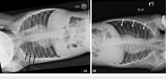
- Pulmonary Conditions:This position is valuable for assessing pleural effusions, which are collections of fluid in the pleural space surrounding the lungs. By utilizing this positioning, the fluid shifts to the dependent portion of the pleural space, allowing for its visualization on chest radiographs.
Additionally, this position facilitates the detection of pneumothorax, a condition where air accumulates in the pleural space, causing a collapse of the lung.
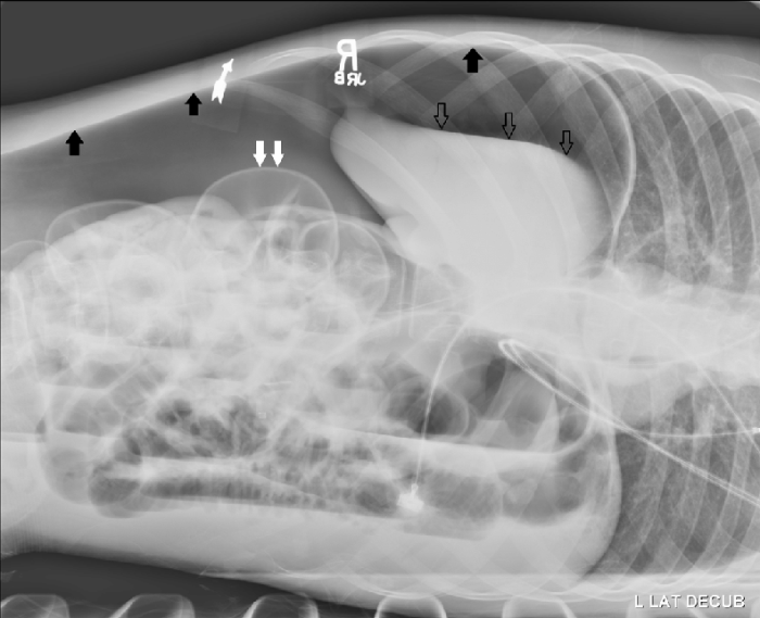
- Abdominal Conditions:In the left lateral decubitus position, free air or fluid within the abdomen will gravitate to the most non-dependent portion, which is the right upper quadrant. This positioning aids in diagnosing conditions like pneumoperitoneum (presence of free air in the abdomen) and ascites (accumulation of fluid in the peritoneal cavity).
- Pelvic Conditions:This position is commonly employed in female patients to assess pelvic structures. It allows for better visualization of the uterus, ovaries, and fallopian tubes. It also assists in diagnosing conditions like uterine prolapse and ovarian cysts.
- Monitoring of Drainage Tubes:In cases where drainage tubes are inserted into the pleural or peritoneal cavities, the left lateral decubitus position can be used to monitor their placement and ensure proper drainage.
FAQ Corner
What is the purpose of a left lateral decubitus position x-ray?
This specialized x-ray examination provides detailed visualization of the heart, lungs, and mediastinum, aiding in the diagnosis and monitoring of various cardiopulmonary conditions.
How is the patient positioned for a left lateral decubitus position x-ray?
The patient lies on their left side, with their left arm raised above their head and their right arm resting comfortably by their side. This positioning allows for optimal visualization of the target anatomical structures.
What are some common pathological findings that can be detected using a left lateral decubitus position x-ray?
This technique can reveal a wide range of abnormalities, including pleural effusions, pneumothorax, cardiomegaly, and mediastinal masses.