Dive into the fascinating world of animal cell coloring! This guide, the animal cell coloring answer key, will unlock the secrets of animal cell structures, making learning both engaging and informative.
As you embark on this journey, you’ll discover the significance of animal cells, unravel the functions of their organelles, and explore the techniques used to color them. Prepare to be amazed by the insights gained through animal cell coloring, its applications in research and education, and the troubleshooting tips to ensure success.
Introduction

An animal cell is the basic unit of life for all animals. It is responsible for all of the activities that an animal needs to survive, such as metabolism, reproduction, and growth. Animal cells are very complex and contain a variety of organelles, each of which has a specific function.
Coloring animal cells is a great way to learn about their different parts and how they work together. It can also be a fun and relaxing activity. There are many different ways to color animal cells, and you can use any colors you like.
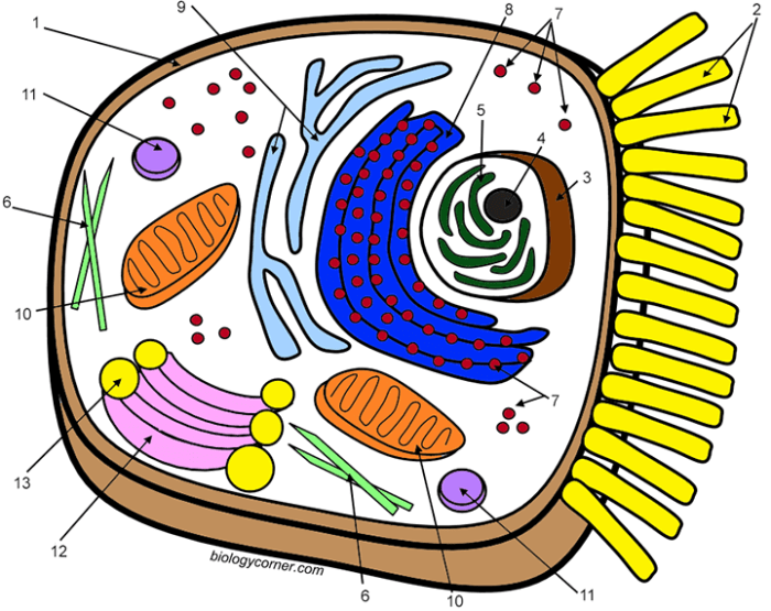
Animal Cell Coloring Answer Key
An animal cell coloring answer key is a guide that shows you how to color the different parts of an animal cell. This can be helpful if you are new to coloring animal cells or if you want to make sure that you are coloring them correctly.
Key Components of an Animal Cell

Animal cells are the fundamental building blocks of multicellular animals. They are responsible for carrying out a wide range of functions, from metabolism and growth to reproduction and movement. To perform these functions, animal cells contain a variety of organelles, each with its own specific role.
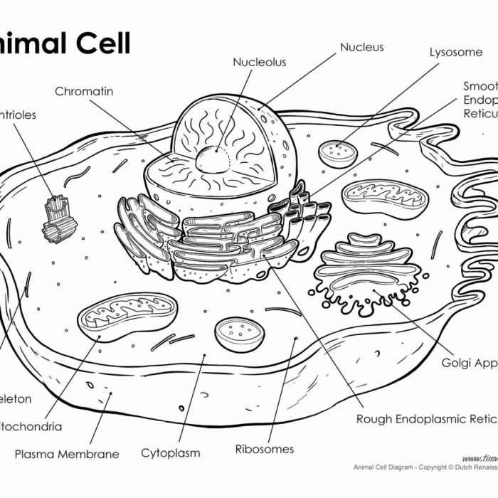
The main organelles of an animal cell include the nucleus, endoplasmic reticulum, Golgi apparatus, mitochondria, ribosomes, lysosomes, and vacuoles. Each of these organelles has a unique structure and function, and they work together to maintain the cell’s homeostasis and carry out its various activities.
Nucleus
The nucleus is the control center of the cell. It contains the cell’s DNA, which is organized into chromosomes. The nucleus also contains the nucleolus, which is responsible for producing ribosomes.
Endoplasmic Reticulum
The endoplasmic reticulum is a network of membranes that folds and transports proteins. There are two types of endoplasmic reticulum: rough endoplasmic reticulum and smooth endoplasmic reticulum. Rough endoplasmic reticulum has ribosomes attached to its surface, while smooth endoplasmic reticulum does not.
Golgi Apparatus
The Golgi apparatus is a stack of flattened membranes that modifies and packages proteins. It also helps to transport proteins to their final destination.
Mitochondria
Mitochondria are the powerhouses of the cell. They produce ATP, which is the cell’s main source of energy.
If you’re looking for the answers to your animal cell coloring assignment, we’ve got you covered. And while you’re here, why not check out our life cycle of stars worksheet ? It’s a great way to learn about the fascinating journey of stars from birth to death.
And who knows, it might even inspire you to become an astronomer! But don’t forget to come back and check those animal cell coloring answers.
Ribosomes
Ribosomes are small organelles that are responsible for protein synthesis. They are found in the cytoplasm and on the surface of the rough endoplasmic reticulum.
Lysosomes, Animal cell coloring answer key
Lysosomes are small organelles that contain digestive enzymes. They help to break down waste products and cellular debris.
Vacuoles
Vacuoles are membrane-bound sacs that store materials. They can store water, food, waste products, and other materials.
The organelles of an animal cell work together to maintain the cell’s homeostasis and carry out its various activities. For example, the nucleus controls the cell’s activities, the endoplasmic reticulum folds and transports proteins, the Golgi apparatus modifies and packages proteins, the mitochondria produce energy, the ribosomes synthesize proteins, the lysosomes break down waste products, and the vacuoles store materials.
Coloring Methods and Techniques
Coloring animal cells is a crucial step in microscopy, allowing researchers to visualize and distinguish different cellular components. Various methods and techniques are employed for this purpose, each with its advantages and disadvantages.
The choice of coloring method depends on factors such as the specific cellular structures to be visualized, the desired level of detail, and the compatibility with subsequent experimental procedures.
Staining Techniques
Staining techniques involve the use of dyes or stains to impart color to specific cellular components. These techniques can be categorized into two main types: vital staining and non-vital staining.
- Vital staining: Involves the use of dyes that can penetrate living cells without causing harm. These dyes are typically used to stain specific organelles or cellular processes.
- Non-vital staining: Employs dyes that can only penetrate cells that have lost their membrane integrity. These dyes are commonly used to stain dead cells or to visualize specific cellular structures after fixation.
Immunofluorescence
Immunofluorescence is a technique that utilizes antibodies labeled with fluorescent dyes to specifically bind to target proteins within cells. This method allows for the visualization of specific proteins or protein complexes within the cell.
Immunofluorescence involves several steps, including antibody incubation, washing, and fluorescence microscopy. The choice of fluorescent dye depends on the excitation and emission wavelengths required for visualization.
Confocal Microscopy
Confocal microscopy is a technique that combines optical sectioning with fluorescence microscopy. It allows for the visualization of specific cellular structures in three dimensions by capturing images at different depths within the cell.
Confocal microscopy uses a laser to scan the specimen, and the emitted fluorescence is collected through a pinhole to eliminate out-of-focus light. This results in high-resolution images with reduced background noise.
Electron Microscopy
Electron microscopy is a technique that uses a beam of electrons to visualize the ultrastructure of cells and tissues. It provides much higher resolution than light microscopy, allowing for the visualization of individual molecules and organelles.
Electron microscopy requires specialized equipment and sample preparation techniques. It is commonly used in research to study the detailed structure of cells and tissues.
Interpretation of Animal Cell Coloring Results
Interpreting the results of animal cell coloring involves analyzing the patterns and colors observed in the stained cells. These patterns and colors provide insights into the cell’s structure, function, and composition.
When examining stained animal cells, it’s crucial to consider the specific staining technique used. Different staining methods target different cellular components, such as the nucleus, cytoplasm, or organelles. Understanding the specific staining method used will guide the interpretation of the observed patterns and colors.
Common Patterns and Colors
Some common patterns and colors observed in animal cell coloring include:
- Nuclear staining:Nuclei are typically stained with basic dyes, such as hematoxylin, which give them a blue or purple color. The size, shape, and chromatin distribution within the nucleus can provide information about the cell’s stage in the cell cycle and its overall health.
- Cytoplasmic staining:Cytoplasm can be stained with acidic dyes, such as eosin, which gives it a pink or red color. The distribution of organelles and other cytoplasmic components can provide insights into the cell’s metabolic activity and function.
- Organelle staining:Specific organelles, such as mitochondria or Golgi bodies, can be stained using specialized dyes. These dyes allow researchers to visualize the distribution and abundance of these organelles within the cell.
Identifying Cell Types
The patterns and colors observed in animal cell coloring can be used to identify different types of animal cells. For example:
- Neurons:Neurons have a large, centrally located nucleus and long, branched extensions called axons and dendrites. They are typically stained with basic dyes, which give them a blue or purple color.
- Muscle cells:Muscle cells have a long, cylindrical shape and contain multiple nuclei. They are typically stained with eosin, which gives them a pink or red color.
- Epithelial cells:Epithelial cells form a continuous layer of cells that lines the surfaces of the body. They are typically stained with hematoxylin and eosin, which gives them a blue or purple nucleus and a pink or red cytoplasm.
Applications of Animal Cell Coloring: Animal Cell Coloring Answer Key
Animal cell coloring has a wide range of applications in scientific research, education, and diagnostics. It provides valuable insights into cell structure, function, and behavior, facilitating advancements in various fields.
In Research
In research, animal cell coloring enables detailed visualization and analysis of cell components. It helps identify specific proteins, organelles, and structures, allowing researchers to study their localization, distribution, and interactions. This information is crucial for understanding cell biology, disease mechanisms, and potential therapeutic interventions.
In Education
Animal cell coloring is a powerful educational tool. It provides students with a hands-on and engaging way to learn about cell structure and function. By coloring different cell components, students can reinforce their understanding of cell biology and develop a deeper appreciation for the complexity and diversity of life.
In Diagnostics
Animal cell coloring plays a significant role in diagnostics. It allows pathologists and medical professionals to identify abnormal cells and tissues, aiding in the diagnosis of diseases such as cancer and genetic disorders. By staining specific cell components, such as nuclei or chromosomes, clinicians can detect abnormalities that may indicate disease progression or provide prognostic information.
Troubleshooting and Resources
Encountering problems during animal cell coloring is not uncommon. Identifying these issues and finding effective solutions can enhance the accuracy and efficiency of your coloring process.
Here are some common issues and troubleshooting tips to consider:
Color Bleeding
When colors spread beyond the intended areas, causing blurred or imprecise lines, it’s known as color bleeding. This can occur due to factors like using too much water or applying excessive pressure while coloring.
- Use less water to prevent the colors from becoming diluted and bleeding.
- Apply a light touch while coloring to avoid pressing too hard and causing the colors to spread.
Uneven Coloring
If some areas of the animal cell appear darker or lighter than others, it may indicate uneven coloring. This can happen when the color is not applied consistently or if the coloring tools are not properly cleaned.
- Ensure thorough mixing of the colors to achieve a uniform consistency.
- Clean the brushes or other coloring tools regularly to prevent color contamination and uneven application.
Resources for Further Learning and Support
To enhance your understanding and skills in animal cell coloring, consider these valuable resources:
- Online tutorials and videos: Numerous platforms offer comprehensive guides and demonstrations on animal cell coloring techniques.
- Books and articles: Refer to reputable publications that provide detailed explanations and illustrations of animal cell structures and coloring methods.
- Online forums and communities: Engage with fellow enthusiasts and experts in animal cell coloring to share experiences, ask questions, and gain insights.
Top FAQs
What is the purpose of coloring animal cells?
Coloring animal cells allows for the visualization and differentiation of various organelles, aiding in the understanding of their functions and relationships within the cell.
What are the common methods used for coloring animal cells?
Animal cells can be colored using stains, dyes, or fluorescent markers, each with its own advantages and disadvantages.
How can animal cell coloring be used in research?
Animal cell coloring is employed in research to study cell structure, dynamics, and interactions, providing valuable insights into cellular processes and diseases.